09/13/2013
RESPONSES / COMMENTS - (CLINICAL) - PART 1A
RE: Recurring Lesions
From: Ed Cohen, DPM
I have seen about 10 of these lesions in the last 35 years. They are usually on the second toe and many times bilateral. I have had great success doing an MIS partial plantar proximal phalangeal head resections, and occasionally an MIS proximal phalangeal head resection. As far as I know, everyone of these surgeries has been successful in getting rid of these lesions.
Ed Cohen, DPM, Gulfport, MS, ECohen1344@aol.com
09/10/2013
RESPONSES / COMMENTS - (CLINICAL) - PART 1C
RE: Gangrene S/P Cast Complication
From: Khurram Khan, DPM
What type of procedure was performed? Is it a possible compartment syndrome after the surgery? Sickle cell? Vasculitis? All these need to be worked up.
1- Given the proximal aspect of the incision site, refer the patient to vascular to assess the PT artery - it may have been injured in the surgery both at the ankle and its branches in the midfoot.
2- PT nerve block for sympathetic blockade.
3- Nitro paste/patch for vasodilation in the area.
4 - Warm compress behind the knee.
5 - An anecdotal suggestion would be to use Metanx to help NO production.
Khurram Khan, DPM, NY, NY, khankhurram@hotmail.com
09/10/2013
RESPONSES / COMMENTS - (CLINICAL) - PART 1B
RE: Gangrene S/P Cast Complication
From: Wm. Barry Turner, BSN, DPM
It may already be too late, but I would try to instigate the use of oral and topical vasodilators. Using HBOT is a great idea, but keep in mind that O2 is a vasoconstrictor. The blood fluid will be richer in oxygen. If you couple the oxygen therapy with a vasodilator, you will see a much quicker and maximized response. I do not like the response quoted from the ortho doctor. I hope his licensing organization is aware of his callous concern for this patient. Topically, I would rub in 1/2 inch of nitroglycerin pasted to the affected foot's arch, tid. Hold for SBP under 100mm. Discuss with the patient's PMD about using oral medication, like Procardia.
My question to the patient's parent, "how did the child tolerate the significant pain that would accompany this travesty?"
Wm. Barry Turner, BSN, DPM, Royston, GA, claret32853@ymail.com
08/16/2013
RESPONSES / COMMENTS - (CLINICAL) - PART 1
RE: Severe Heel Pain After Plantar Fasciotomy (Mark Aldrich, DPM)
From: David Zuckerman, DPM
It is very nice that we have additional treatment modalities such as RF and/or lasers to treat post-surgical pain and inflammation. Lasers can heal nerve problems such as neuropathy and more. I stress that the most important aspect of dealing with a complication is very simple - find out what the diagnosis is, and just don't throw a laser or RF at the problem. I am not saying that you can't help this specific case with a laser, but you won't help the patient without a specific diagnosis. Do a complete blood work-up. I have seen five cases of anklyosing spondylitis post-ESWT.
David Zuckerman, DPM, Cherry Hill, NJ, footcare@comcast.net
06/14/2013
RESPONSES / COMMENTS - (CLINICAL) - PART 1B
RE: Recurrent Ganglion Cysts
From: Steven J. Kaniadakis, DPM
Typically, I inject Depomedrol just proximal to the post-surgical wound. According to O.A. Mercado's videos, and other various reference textbooks, a "few drops of Decadron" intra-operatively is another technique. The post related to using "non-absorbable suture and to heavily cauterize it." I suspect that this might be causing further perpetuating aggravating factors such as the irritation of the surrounding structures, and causing the cycle to repeat from repetitive irritation of remnant sutures, perhaps overzealous necrotic cauterized tissue.
I would have selected an absorbable type suture to help nature maintain a smooth natural gliding action. If it's emerging from a (synovial) joint, then there may be iatragenic causes that are inducing tendon sheath ganglionic cyst(s). Finally, a firm compression post-op dressing helps with a mixture of long and short-acting corticosteroids.
Steven J. Kaniadakis, DPM, St. Petersburg, FL, stevenkdpm@yahoo.com
06/11/2013
RESPONSES / COMMENTS - (CLINICAL) - PART 1B
RE: Recurring Skin Lesions
From: Dennis Shavelson, DPM
This is a case of biomechanical surgical iatrogeny. These are not recurrent lesions as Name Withheld has labeled them; they are transfer lesions. The surgeon has stripped the distal segment of the 3rd digits in question from being functional in closed chain. The patient has morphed mechanically to a compensatory equilibrium via Wolf’s and Davis’ laws. That is giving a new location of the digits weight-bearing purpose which they are not biologically capable of assuming, resulting in this pathological dermatological reaction.
Perform a biomechanical evaluation of the digits, beginning with functional foot typing, and take a biopsy of the lesion. Send the specimen to a pathology lab (such as Bako) that can give you a biomechanical microscopic reading of the slide. Utilizing that data, develop a non-operative and/or operative plan of care to resolve this painful and disabling problem.
Disclosure: I am a consultant to Bako Labs.
Dennis Shavelson, DPM, NY, NY, drsha@foothelpers.com
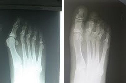
06/11/2013
RESPONSES / COMMENTS - (CLINICAL) - PART 1A
RE: Recurring Skin Lesions
From: Gino Scartozzi, DPM
Radiographs would be helpful to determine underlying factors contributory to the redevelopment of these lesions. However, I suggest it is possible that the third metatarsals are elongated relative to the adjacent second metatarsals. Such elongated metatarsal positions may be contributory to such biomechanically-induced lesions on the plantar aspect of the third toes seen despite toe arthroplasties being previously performed.
If the third metatarsals are elongated relative to the adjacent metatarsals, I suggest orthotic management, or shortening osteotomy of the third metatarsals if orthotics fail.
Gino Scartozzi, DPM, New Hyde Park, NY, Gsdpm@aol.com
06/10/2013
RESPONSES / COMMENTS - (CLINICAL) - PART 1B
RE: Recurring Skin Lesions
From: John Scheffel, DPM
It's hard to tell without x-rays, but I wonder if the initial surgeon performed a flexor tendon transfer instead of just a tenotomy? I had a similar case in which the proximal phalanx was causing a similar problem. The patient had the procedure done years earlier, and all that was left of the proximal phalanx was its base, which was sitting distal-plantar to the met head.
Excision of the base of the proximal phalanx with syndactylization to the adjacent digit addressed her pain.
John Scheffel, DPM, Worcester, MA, jscheffel@townisp.com
06/10/2013
RESPONSES / COMMENTS - (CLINICAL) - PART 1A
RE: Recurring Skin Lesions
From: Elliot Udell, DPM, Jon Purdy, DPM
I have seen many similar lesions. I like to keep things simple and work upward from there. With an unknown skin lesion step-one is biopsy and send the specimen to a dermatopathologist. If benign, it’s time to break the cycle of formation. I simply apply a high potency steroid under occlusion. It will resolve and may need to be performed from time to time. Many times, never again.
Jon Purdy, DPM, New Iberia, LA, podiatrist@mindspring.com
This patient might benefit from gait pressure studies in order to determine if there is excess pressure beneath those areas during gait. There are many products available that can perform this study. The one we use is the inexpensive carbon paper based device. The person walks over it and it determines where there is excessive pressure during ambulation. There are computer-based electrical plates that do the same thing. If it is determined that this is the case, then you can address the problem biomechanically and design a set of orthoses to correct the problem. Such information may also help if you intend to address the problem surgically.
Elliot Udell, DPM, Hicksville, NY, Elliotu@aol.com
05/27/2013
RESPONSES / COMMENTS - (CLINICAL) - PART 1
RE: Non-Union of the Hallux IPJ
From: Jefferson J. Mennuti, DPM
If you are only considering surgical correction for the non-union, then you would have to remove the implant, pack the deficit with auto-graft marrow, which you can get from the calcaneus, and use 2 compression Nitanol staples, one medial, one lateral. If you feel there would be lack of cortical purchase, consider a locking plate rather than the staples.
Jefferson J. Mennuti, DPM, Orange City, FL, dr.mennuti@gmail.com
05/24/2013
RESPONSES / COMMENTS - (CLINICAL) - PART 1
RE: Non-Union of the Hallux IPJ
From: Aidan Nguyen, DPM
Cases like this exemplify the overuse of surgical innovation while neglecting basics of fusion principles, i.e. thorough joint curettage and dynamic compression. I wouldn’t revise an asymptomatic (non-painful) nonunion, especially at the toe level, although your concern of cortical fracture is valid.
Regarding your surgical query, I would remove the implant, re-prep the fusion site, and use either a 3.5 cortical or 4.0 cannulated screw, compressing the joint from the toe distally. Using a bone graft to maintain length post-revision is worth considering, although it would complicate the compression screw placement process.
Aidan Nguyen, DPM, Yuba City, CA, nguyena2@sutterhealth.org
05/18/2013
RESPONSES / COMMENTS - (CLINICAL) - PART 1B
RE: Painful Bunion in a 12 Year Old
From: Name Withheld
Thank you to those who gave me your input. I did not provide an extensive biomechanical exam with my first presentation. This patient has at least 10 degrees of ankle dorsiflexion with her knee extended or flexed. Her hamstrings are not tight. She has symmetrical hip motion, more with no internal position. There is no internal tibial torsion. She does not have ligamentous laxity. Her first ray is stable on exam. She is only moderately pronated in stance and gait. Her lateral x-ray does not have an anterior break in the cyma line. There is no Kirby's sign. There is no elevation of 1st ray (Seeberg's index).
She has noticed the bunions for several years, and has had moderate aching in the joint for...
Editor's note: This extended-length letter can be read here
05/16/2013
RESPONSES / COMMENTS - (CLINICAL) - PART 1B
RE: Painful Bunion in a 12 Year Old
From: Gary W Docks, DPM
The metatarsus adductus and positive metatarsal protrusion factor highly when considering the bunion correction. One thing you did not mention is whether she had a significant equinus, which definitely is causal when you're looking at a juvenile bunion deformity. That being said, you must also be aggressive in choosing your procedure versus being conservative, which likely will result in a recurrence. Remember, the met adductus deformity will increase the IM angle. I therefore recommend the following procedures:
1. Endoscopic gastroc recession 2. Scarf or Austin Bunionectomy with screw fixation. There are still remnants of the growth plate at the base of the lst metatarsal. You could perform a Juvara, but you would have to be careful to avoid the growth plate. Remember to be aggressive and reduce the IM angle or 10 years from now, you'll be looking at a re-do.
Gary W Docks, DPM, Beverly Hills, MI, gwdocks@aol.com
05/11/2013
RESPONSES / COMMENTS - (CLINICAL) - PART 1B
RE: Overlapping 2nd Digit Status Post Bunionectomy
From: Don Peacock, DPM)
I wanted to show a nice correction I got on a bunion deformity with the patient on whom I felt uncomfortable with doing a 1st metatarsal osteotomy. In your case, you would have to remove the fixation in the first metatarsal. This can complicate the redo procedure for the patient. On the elderly patient, I’m going to show I performed a minimally invasive bunionectomy and Akin through and through.
 |
Pre- and Post-op X-Rays, Akin Performed Minimally Invasive |
I used the same incision percutaneously to perform the bunionectomy and the Akin. The interesting result with her x-rays is that...
Editor's note: Dr. Peacock's extended-length letter can be read here.
05/08/2013
RESPONSES / COMMENTS - (CLINICAL) - PART 1B
RE: Chronic First MPJ Pain (Don Peacock, DPM)
From: Sloan Gordon, DPM
Dr. Peacock, your x-rays are most impressive. You plantarflexed the first ray below the dorsal cortex, which will off-load the first ray, and you removed the 1st MTPJ exostosis just perfectly. I am not sure how you did this through a 1.5 cm incision, unless this was MIS. I guess we Texans like things big, including our incisions!
If this was MIS, I would be concerned about a non-fixated metatarsal osteotomy along with the gastoc lengthening. It seems like taking a risk for displacement of the osteotomy. But, the pre- and post-operative films are impressive. If this was MIS, why not drive a percutaneous 0.062" Kirschner wire through the osteotomy? I am certainly not faulting your technique!
Sloan Gordon, DPM, Houston, TX, sgordondoc@sbcglobal.net
05/08/2013
RESPONSES / COMMENTS - (CLINICAL) - PART 1A
RE: Chronic First MPJ Pain (Don Peacock, DPM)
From: Charles Morelli, DPM
Not having seen this patient and basing these comment solely on the x-ray, I respectfully submit that this patient's condition was never addressed properly. Pre-op, there is an elevated first met and despite efforts to plantarflex the head, it is still dorsiflexed. Pre-op, there is a dorsal exostosis, and I would be surprised if there is any increase in hallux dorsiflexion. The dorsal bump was not removed aggressively enough and removing part of the dorsal aspect of the base of the proximal phalanx could have also been considered.
If you make a template and find the CORA, I believe you will see that this patient could have benefited from a Cotton procedure that would have required a graft and possible plate; depending on graft chosen. I don't want to, nor will I, get into a debate about MIS vs. open, but this case required more that a 1.5 cm incision and needed to be treated much more aggressively.
Charles Morelli, DPM, Mamaroneck, NY, podiodoc@gmail.com
05/07/2013
RESPONSES / COMMENTS - (CLINICAL) - PART 1
RE: Pedal Reaction to Doxil
From: Elliot Udell, DPM
This patient presented with a pedal skin reaction to Doxil, which is a chemotherapy drug. Skin reactions are a known side-effect of this pharmaceutical. The skin reaction developed within days of taking the drug.
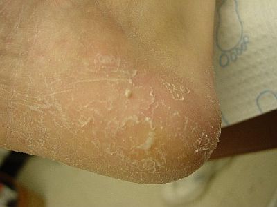 |
Skin Reaction to Doxil |
Initially, the redness was far more severe and her oncologist controlled it with topical steroids. The scaling and cracking remains, and we are managing it with a 30% urea-based cream in addition to the topical steroid.
Elliot Udell, DPM, Hicksville, NY, Elliotu@aol.com
05/04/2013
RESPONSES / COMMENTS - (CLINICAL) - PART 1B
RE: Post-Keller Complication (Gregory Caringi, DPM)
From: Don Peacock, DPM
Unfortunately, this case is a result of an inappropriate procedure performed on a 54 year-old patient. Keller procedures should be used with caution and have a significant history of complications. In Keller’s original paper, he discussed that he also performed an extensor hallucis longus lengthening. This is overlooked by some surgeons. Kellers are intended for use in the elderly.
Two of the most significant deformities resulting from Keller procedures are...
Editor's note: Dr. Peacock's extended-length letter can be read here.
05/02/2013
RESPONSES / COMMENTS - (CLINICAL) - PART 1
RE: Foreign Body Granuloma? (David Kahan, DPM)
From: Neil Levin, DPM
Avulse the entire nail. Debride the granulomatous tissue and send it all for biopsy. Treat it post-operatively as you would any avulsion until you have path results.
Neil Levin, DPM, Sycamore, IL, DRFEET1@aol.com
05/01/2013
RESPONSES / COMMENTS - (CLINICAL) - PART 1
RE: Chronic Onychomadesis (Joseph Reynolds, DPM)
From: Neil H Hecht, DPM
Distal disruption of nail plate to nail bed is termed onycholysis. I know of no acceptable surgical procedure to “tack down” this non-living protein (keratin) nail plate to a living nail bed. This doesn’t make any sense to me on a physiologic basis.
Proximal disruption of the nail plate to the nail bed is termed onychomadesis. In case any of the readers remember Dr. Philip Gardner (CCPM in the 1970s), the nail groove callus is termed onychophosis. I use onychauxis to describe a thickened (hypertrophic) nail and onychogryphyosis to describe a grossly distorted nail (“claw nail”). Both would be examples of onychodystrophy.
Neil H Hecht, DPM, Tarzana, CA, drhecht@drneilhecht.com
04/25/2013
RESPONSES / COMMENTS - (CLINICAL) - PART 1B
RE: Post-Op Hallux Varus (Kel Sherkin, DPM)
From: Robert Bijak, DPM, Jeffrey Kass, DPM
Simply correct the PASA with a Reverdin.
Robert Bijak, DPM, Clarence Center, NY, rbijak@aol.com
If there is no pain, then of course one doesn't have to repair it. The patient does have a complaint and if you to chose to address it, you may want to consider the Arthrex mini tight rope. I would like to add, a podiatrist could have had the same outcome.
Jeffrey Kass, DPM, Forest Hills, NY jeffckass@aol.com
04/24/2013
RESPONSES / COMMENTS - (CLINICAL) - PART 1B
RE: Post-Op Hallux Varus (Kel Sherkin, DPM)
From: Ira Weiner, DPM
Based upon the x-ray findings and history, I feel a closing Aiken osteotomy is in order. Since she states that the MPJ has a pain-free ROM, I would not address that. It appears she does not have the classic "staked" metatarsal. If a fusion procedure is warranted in the future, there is plenty of good bone left to support that procedure using any fixation method of choice.
As a side note, the question of cosmesis only is brought up. I feel if she has difficulty finding the shoes she wants, it becomes her call as to whether or not she wants this corrected. I don't feel there is a single surgeon among us who does not strive for good cosmesis in an elective procedure. Salvage procedures are a little different. If the patient is unhappy with the way the procedure looks, then we have a duty to correct the problem if it's something we are able to do.
Ira Weiner, DPM, Las Vegas, NV, vegasfootdoc2005@yahoo.com
04/20/2013
RESPONSES / COMMENTS - (CLINICAL) - PART 1B
RE: Bacterial Infection of Nail (Jay Kerner, DPM)
From: Martin V. Sloan, DPM
I had similar reported findings after I submitted a nail specimen to a major pathology lab to rule out onychomycosis. The report said "bacterial overgrowth with no fungal elements seen," and identified this same staph organism, Staph hemolyticus. The patient acquired her condition at a nail spa and wanted it resolved. So I put her on an appropriate anti-staph antibiotic (not Cipro) for a time, and the condition resolved. I don't know whether it was a colonization or an infection, but that debate could be extended to the dreaded fungal toenail as well.
Martin V. Sloan, DPM, Rowlett, TX, martinsloan@me.com
04/19/2013
RESPONSES / COMMENTS - (CLINICAL) - PART 1B
RE: Wart Covered Foot (David Kahan, DPM)
From: Philbert Kuo, DPM, Brian Kiel, DPM
This looks like a good case for the Panacos procedure. David Secord, DPM has discussed this procedure and Peter Bregman, DPM has written about it.
Philbert Kuo, DPM, Chesapeake, VA, philbertkuo@gmail.com
With patients like this I have successfully treated the condition with once daily applications of 10% formalin solution (formerly Lazerformalyde) which can be made by your local pharmacy and 1200 mg of cimetidine (Tagamet) given in a single or divided dose. I see them after 3 weeks for debridement and then every 6 weeks. I have seen this be successful in a matter of weeks, but usually it takes 4-8 months.
Brian Kiel, DPM, Memphis, TN, Footdok4@gmail.com
04/19/2013
RESPONSES / COMMENTS - (CLINICAL) - PART 1A
RE: Wart Covered Foot (David Kahan, DPM)
From: David Hettinger, DPM, Elliot Udell, DPM
I would ABSOLUTELY get him on a regimen of cimetidine 400mg tid for twelve to sixteen weeks. (JAPMA, November 1, 1995 vol. 85 no. 11 717-718).
David Hettinger, DPM, Wheaton, IL, davidhett@msn.com
I have often seen cases of verrucae, especially mosaic verrucae spread all over the bottom of a persons' foot. It is rare but possible to see it spread to the hands. There might be more going on with this patient than verrucae. You might wish to do a "scoop out" procedure on one or two of the warts and send them to a dermatopathology lab in order to be certain that you are indeed treating verrucae. You might also want to send this patient to an immunologist in order to rule out the possibility of a deficiency in his immune system.
Elliot Udell, DPM, Hicksville, NY, Elliotu@aol.com