09/04/2013
RESPONSES / COMMENTS (CLINICAL) - PART 1A
RE: Bunion with Overlapping Toe
From: Michael B. DeBrule, DPM
Co, et al. looked at different interventions for 2nd toe transverse plane deformity in JFAS back in 2006. Syndacylization yielded the best alignment results compared to lateral anchor suture, flexor transfer, flexor plate repair etc. Therefore, I suggest: sew the 2nd and 3rd together, along with hammertoe corrections; 1st MTPJ athrodesis (more predictable toe alignment than with an Austin); and maybe a Weil 2nd osteotomy (depending on met length on x-rays).
On the other hand, the clinical picture seems more appropriate for an external metatarsal bar attached to an extra depth/custom shoe, custom foot orthotics, lift to correct limb length, periodic paring of the hyperkeratosis, or something conservative here. Consider discussing your proposed surgical intervention with the primary care doctor, physical therapist, and family. Would surgery truly improve her quality of life and help her walk better? Or would the patient be more likely to fall and sustain a hip fracture from wearing a CAM boot?
Michael B. DeBrule, DPM, Richfield, MN, innovativefootcare@gmail.com
09/04/2013
RESPONSES / COMMENTS (CLINICAL) - PART 1B
RE: Bunion With Overlapping 2nd Toe
From: Ed Cohen, DPM
A good point was raised about patient selection. A person with a drug or alcohol problem should be carefully screened and you might want to limit these type of patients to the more simple surgical procedures. It may be prudent not to take this case.
Amputation of the second toe should be avoided, as it will leave a space between the first and third toes, which will eventually cause the lateral deviation of the big toe and recurrence of the bunion. Also, an isolated second metatarsal head resection will ruin the...
Editor's note: Dr. Cohen's extended-length letter can be read here.
09/02/2013
RESPONSES / COMMENTS (CLINICAL) - PART 1B
RE: Bunion With Overlapping 2nd Toe
From: Tip Sullivan, DPM, Michael J Marcus, DPM
Do you want to do foot surgery on a middle aged, non-compliant person who just happens to be an ex-addict? Is surgery the right thing to do? There is no doubt that surgical correction of this foot deformity is possible without the patient losing a toe. Whether you fuse the 1st MTPJ and syndactylize the second or do some other more proximal procedure, the question to ask here is one of patient selection for surgical intervention. If this unfortunate young gunshot victim is in a nursing home environment and is not walking much, why not get her some custom-molded shoes? If you do choose a surgical route for this patient, my advice would be “protect her from herself”— AK cast, knee flexed, toe pointed — and in a wheel chair until bone union.
Tip Sullivan, DPM, Jackson, MS, tsdefeet@msfootcenter.net
If symptomatic, I would perform an arthrodesis to the first MPJ together with a Weil osteotomy on the 2nd, arthrotomy/capsulotomy, or a plantar plate repair on the 2nd MPJ with a hammertoe fusion. These deformities are rigid secondary to neurologic etiology - gun shot/nerve injury. Nothing less than an arthrodesis would maintain the correction.
Michael J Marcus, DPM, Montebello/Irvine, CA, ftmed@aol.com
07/29/2013
RESPONSES / COMMENTS (CLINICAL) - PART 1A (CLOSED)
RE: Chronic Pruritic Lesion (Arthur Gudeon, DPM)
From: Don Steinfeld, DPM
That looks like lichen simplex chronicus to me. Originally a pruritic lesion, the skin becomes indurated with exaggerated skin lines due to repetitive excoriation. This is an example of neuro-dermatitis. Try an Unna boot to physically prevent access to the site.
Don Steinfeld, DPM, Farmingdale, NJ, footdrdon@aol.com
07/29/2013
RESPONSES / COMMENTS (CLINICAL) - PART 1B (CLOSED)
RE: Chronic Pruritic Lesion (Arthur Gudeon, DPM)
From: Art Gudeon, DPM
Thanks for your responses; they’re much appreciated. As it turns out, Bryan Markinson, DPM, my “go-to podiatric dermatologist,” agreed with the consensus that the condition was NOT psoriasis, but lichen simplex chronicus. He advised treating this condition with clobetasol 0.05% bid (basically the same potency as the betamethasone dipropionate 0.05% ung., bid, that I’d already prescribed). I can alter the Rx later if necessary, and also add Benadryl prn (although she says the pruritis is minimal right now).
Just to clarify the biopsy situation - the reason she preferred not to have it on her first visit was due to a swimming competition “final” later that day, but said she’d want it if the response wasn’t proving satisfactory over a “reasonable” period of time. She was aware she’d have to D/C treatment 48-72 hours prior to the biopsy if so.
Art Gudeon, DPM, Rego Park, NY, afootdoc@hotmail.com
12/10/2012
RESPONSES / COMMENTS (CLINICAL) - PART 1B
RE: A1c and Elective Foot Surgery (Allen Jacobs, DPM)
From: Tip Sullivan, DPM
Dr. Jacobs has opened a very appropriate topic for discussion. I am not aware of any papers that specifically relate A1c to foot surgery success, elective or otherwise. The first thing that I asked myself after considering his question was: What is “elective” foot surgery? Certainly, we all agree that the current trend in “cosmetic” foot surgery is “elective.” I think the line one draws between elective surgery and necessary surgery can get grey.
Perhaps the terms emergent and non-emergent surgery would be...
Editor's note: Dr. Sullivan's extended-length letter can be read here.
12/08/2012
RESPONSES / COMMENTS (CLINICAL) - PART 1 A
RE: A1c and Elective Foot Surgery (Allen Jacobs, DPM)
From: Pat Caputo, DPM
We all know that the hemoglobin A1C test measures the average blood glucose control for the past 2 to 3 months and is a good general measure of glycemic control during that period. Due to the nature and definition of glycosylated hemoglobin, it only needs to be performed 3 times per year. I am not so sure it should be considered or classified as standard of care to order a hemoglobin A1c prior to all elective foot surgery, unless it hasn’t been checked in over 3 months. It is, however, a very useful tool in predicting increased risk. Studies have shown that “Elevated pre-operative hemoglobin A1c level is predictive of adverse events after coronary artery bypass surgery” (Halkos, M et al., Journal of Thoracic and Cardiovascular Surgery. 2008. 136(3) 631-640.)
The surgeon has to determine how extensive the planned surgery is and measure all other co-morbidities and factors (obesity, smoking, patient compliance, etc.). A HgA1c level of >9% represents an obvious high level of risk that I wouldn’t want to have my diabetic patient exposed to. A target A1c of under 7% is the most appropriate. As the A1c increases, so does the risk of complications. Like most everything we do, your medical decision is how much risk is worth the benefit.
Pat Caputo, DPM, Holmdel, NJ, capstops@aol.com
12/08/2012
RESPONSES / COMMENTS (CLINICAL) - PART 1B
RE: A1c and Elective Foot Surgery (Allen Jacobs, DPM)
From: Brian Crispell, DPM, Robert Wunderlich, DPM
Our hospital just covered this topic at our last department of surgery meeting on Tuesday December 4th. The chairman of surgery at Lankenau Hospital, Dr. Scott Goldman, advised that nothing above 7.5 should be allowed for elective surgery.
Brian Crispell, DPM, Ardmore, PA, bdcrispell@hotmail.com
Evaluating hemoglobin A1c every 3 months is the standard of care for diabetic patients, whether or not they are scheduled for elective surgery. In my community, the test is typically ordered by the patient's primary care physician (or whoever is actively managing the patient's diabetes). In their pre-operative assessment, if the primary care physician is satisfied that the patient's diabetes is stable and controlled, I wouldn't have a problem performing elective foot surgery (assuming there are no other contraindications to surgery). Generally speaking, these patients will have a recent HbA1c around 7% or less.
Robert Wunderlich, DPM, San Antonio, TX, rwunder@gmail.com
12/04/2012
RESPONSES / COMMENTS (CLINICAL) - PART 1A
RE: Verrucous-Looking Lesions on a Seven Year Old (Dennis Shavelson, DPM)
From: Neil Levin, DPM, Seth J. Steber, DPM
Blunt curette the lesion for biopsy; then laser the base as usual.
Neil Levin, DPM, Sycamore, IL, DRFEET1@aol.com
I use a high-temp cautery pen in the office setting to circumscribe the lesion and then evacuate it with a curette. This leaves the lesion intact for the pathologist to fully evaluate. It also allows preservation of the dermal layer so no scar tissue forms. Closure with sutures is not necessary and not recommended - let it heal by secondary intention. This method works as well as using CO2 and KTP/Yag lasers.
Seth J. Steber, DPM, Lehighton, PA, acpwc@ptd.net
Editor's note: To see the original note and photo, click on the subject line.
12/04/2012
RESPONSES / COMMENTS (CLINICAL) - PART 1B
RE: Verrucous-Looking Lesions on a Seven Year Old (Dennis Shavelson, DPM)
From: Elliot Udell, DPM
If you are not sure of the nature of the lesion and it is too large to do an excisional biopsy, why not do one or two 2 mm. punch biopsies of the lesion and send them to a dermatopathology lab? If it comes back indicating that it is indeed a verruca, then there are many non-surgical choices that you can employ. One treatment we use that is "kid friendly" is called cryoprobe. There are many others.
On the other hand, if the pathology report indicates that the lesion is something other than a benign lesion, then surgical excision might be necessary. If removal would leave a "crater" too large to close, you might need to do a graft, and it would probably be best to call in a plastic surgeon as a consultant.
Elliot Udell, DPM, Hicksville, NY, Elliotu@aol.com
12/03/2012
RESPONSES / COMMENTS (CLINICAL) - PART 1B
RE: 1st MTPJ Fusion or Lapidus for Severe HAV (Gino Scartozzi, DPM)
From: Barry Mullen, DPM
I couldn't agree more with Dr. Scartozzi's logic and sentiment. I simply fail to understand the rationale that supports fusing a salvageable 1st MTP. Proponents of 1st MTP fusion claim excellent functional results post-operatively. Really? What does that mean to a healthy, young, active individual who wants to retain the same activity level post-operatively as they enjoyed pre-operatively? When you have a pre-operative functional 1st MTP, a surgeon's goal should be to retain joint motion, even increase it when possible, not eliminate it.
In this case, while the PASA and IM angles are significantly elevated, a clear joint space exists which indicates...
Editor's note: Dr. Mullen's extended-length letter can be read here.
12/01/2012
RESPONSES / COMMENTS (CLINICAL)
RE: 1st MTPJ Fusion or Lapidus for Severe HAV (Mark Aldrich, DPM)
From: Charles Morelli DPM
Clearly, there is more than either a Lapidus and/or MPJ fusion for a "severe" HAV deformity. For the past few years, I have been utilizing an opening base wedge osteotomy with plate (Arthrex) for this deformity. The procedure has been modified a bit which makes this a remarkably strong and stable construct. Special thanks to my friend and colleague Lester Dennis, DPM for showing me this. This fixation is so stable that my patients are allowed to weight-bear immediately and are placed in a CAM walker for the first 2 weeks, and then allowed to gradually progress to a sneaker.
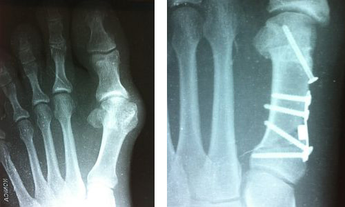 |
Opening Base Wedge Osteotomy with Plate (Arthrex) |
As you can see, one of the screws is crossing the osteotomy at an angle and, by doing this, it is able to purchase all three corticies. This one screw makes this correction very strong and very stable. The medial eminence is also chopped up into appropriate sized pieces and placed in the opening wedge. If desired, you can also add stem cells, Grafton, PRP, Trinity, or any other substance to assist in healing. At conference lectures, everyone states that patients need to be NWB for 4 weeks, and then in a CAM walker for two weeks. You may agree or disagree with this, but immediate weight-bearing is permitted and possible. After doing this procedure in this manner for a number of years now, I have quite a few patients who have done well with it, and never have had a complication.
Charles Morelli DPM, Mamaroneck, NY, podiodoc@gmail.com
11/30/2012
RESPONSES / COMMENTS (CLINICAL) - PART 1A
RE: 1st MTPJ Fusion or Lapidus for Severe HAV (Mark Aldrich, DPM)
From: Gino Scartozzi, DPM
The utilization of a 1st MTPJ fusion for severe hallux abducto valgus deformities WITH the presence of pronounced degenerative joint disease in a young active patient with good bone stock certainly has a surgical indication. In many cases that I have seen, the indication for arthrodesis of the first metatarsal-phalangeal joint is associated more with late Stage II or Stage III joint manifestations in hallux limitus pathology. With these deformities, the intermetatarsal angle deformities tend to be normal to near normal in measurement.
The indications for a Lapidus as a medial column stabilizing procedure is considered with any...
Editor's note: Dr. Scartozzi's extended-length letter can be read here.
11/30/2012
RESPONSES / COMMENTS (CLINICAL) - PART 1B
RE: 1st MTPJ Fusion or Lapidus for Severe HAV (Mark Aldrich, DPM)
From: Edward Cohen, DPM
Fusing a pain-free, non-arthritic MPJ for a high and severe HAV deformity should rarely, if ever, be done as almost all of these bunions can be corrected with a reasonable functional and cosmetic result.
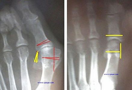 |
Reverdin-Isham-Akin Bunionectomy |
The Reverdin-Isham-Akin bunionectomy is an excellent procedure for correcting this problem, which will occasionally need a closing lateral base wedge osteotomy. The orthopedist from Spain, Carlos Pique Vidal, has a terrific video on his website illustrating this point.
Edward Cohen, DPM, President of AAFAS, ecohen1344@aol.com
11/17/2012
RESPONSES / COMMENTS (CLINICAL) - PART 1 A
RE: Pea-Sized Plantar Fibroma (David Kahan, DPM)
From: Evan F. Meltzer, DPM, Barry Mullen, DPM
I have found that small (<2-3 mm) plantar fibromas often respond to 1 intralesional injection of corticosteroid of your choice. This would seem to be an ideal treatment for your patient.
Evan F. Meltzer, DPM, San Antonio, TX, Evan.Meltzer@va.gov
Are you sure about your diagnosis? While a post-traumatic fibroma is conceivable from a foreign body penetration due to the relative lack of adipose tissue along the plantar surface of the foot and the fascia's anatomic proximity to the dermis, the history of this chief complaint is more highly suggestive of either an epidermal inclusion cyst or foreign body granuloma. In either case, Verapamil is unlikely going to influence this mass. Excision is the treatment of choice. Try pressure dispersing the mass during the athlete's season, and if painful enough to hinder athletic performance, excise it during the off-season.
Barry Mullen, DPM, Hackettstown, NJ, yazy630@aol.com
11/17/2012
RESPONSES / COMMENTS (CLINICAL) - PART 1B
RE: Pea-Sized Plantar Fibroma (David Kahan, DPM)
From: Brad Makimaa, DPM
I clearly would not first assume fibroma. I certainly would not jump to verapamil on a 17 year old. I also would not have a surgical discussion without a proper diagnosis. I would get an MRI. This will give better insight to the diagnosis which can direct the best treatment. I am much more inclined to think more of inclusion cyst or scar tissue as the primary diagnosis. Diagnosis must be established first, i.e. Is this mass attached to the fascia or arising from it? Is there a metal or organic fragment? I assume that an x-ray was done. This vastly changes your surgical planning and discussion with the patient on outcome and post-op scenario.
Brad Makimaa, DPM, Key West, FL, drmak3@comcast.net
10/22/2012
RESPONSES / COMMENTS (CLINICAL) - Part 1A
RE: Chronic Interdigital Maceration (John Scholl, DPM)
From: Robert K Hall, DPM, Robert Bijak, DPM
An effective adjunct to wicking moisture/maceration may be by utilizing Lamb's wool "rope" threaded between the affected toes. While not sterile, topical antibiotics (I prefer cream) or antifungals (like Naftin Gel) may be used. Appropriate bacterial C&S or fungal culture helps ID organism(s) or lack thereof. Use of "prophylactic" antibiotics may trigger a super-infection.
Robert K Hall, DPM, Ft Lauderdale, FL, robertkhalldpm@bellsouth.net
The maceration is due to the biomechanical overexertion of the muscle engines, causing hyperhidrosis, maceration, and infection. A custom foot orthosis, controlling pronation and varus, will restore efficiency, reduce sweating, and allow healing. Orthotics are the answer for most foot problems.
Robert Bijak, DPM, Clarence Center, NY rbijak@aol.com
10/22/2012
RESPONSES / COMMENTS (CLINICAL) - PART 1B
RE: Chronic Interdigital Maceration (John Scholl, DPM)
From: Vito J Rizzo, DPM, Barry Mullen, DPM
Recently, I have begun to offer treatment with laser for this resistant contamination. The half dozen cases performed to date have all resolved with a maximum of 3 treatments.
Vito J Rizzo, DPM, vjrizzo@optonline.net
I'm miffed by responses employing empiric therapy for a common condition that arises from a myriad of etiologies, and amused by the "secret weapon"comment. Presuming the lesion has not arisen from a skin cancer (open wounds not responding to therapy w/in 6 months = biopsy), then determine the etiology BEFORE you treat. This eliminates empiric therapy and provides a basis for evidenced-based medical treatment. In the case of macerated web spaces, how is this accomplished? Well, my "secret weapon" is the Wood's Light = color of florescence = etiology i.e. red = corynea bacteria minitismun = mycins; green = pseudomonas = quinalones; white = psoriasis or...
Editor's note: Dr. Mullen's extended-length letter can be read here.
10/13/2012
RESPONSES / COMMENTS (CLINICAL) - PART 1 A
RE: Chronic Interdigital Maceration (John Scholl, DPM)
From: Michael J Felicetta, DPM, Sloan Gordon, DPM
I have found that this is sometimes related to a gram negative bacterial infection and responds readily to oral antibiosis and daily BID local care with Domeboro's solution, gentamicin cream, and antifungal topical therapy.
Michael J Felicetta, DPM, Toms River, NJ, DrMFoot@aol.com
My secret weapon for this condition is to start a regimen of Metornitazole gel (Metrogel) nightly x 1 month. Often the culprit is corynebacterium mintissimum and this eradicates it well. Of course, I imagine you are controlling sweating and footgear, etc. Of course, if nothing works, biopsy - as is evident in Dr. Carl Solomon's patient.
Sloan Gordon, DPM, Houston, TX, sgordondoc@sbcglobal.net
10/13/2012
RESPONSES / COMMENTS (CLINICAL) - PART 1B
RE: Chronic Interdigital Maceration (John Scholl, DPM)
From: Eric M. Hart, DPM
I have seen benefit to using lamb's wool between the toes in addition to the drying agents you have already used. It can be used as a preventive measure for life. Just make sure that the patient doesn't wrap it circumferentially around the toes.
Also, clearly his problem is more an issue of uncontrolled edema rather than a physical therapist placing gauze between his toes. It will likely recur. I would work with his primary care provider to ensure that diuresis management is maximized in addition to your standard care.
Eric M. Hart, DPM, Bismarck, ND, erichartdpm@gmail.com
10/12/2012
RESPONSES / COMMENTS (CLINICAL) - PART 1A
RE: Chronic Interdigital Maceration (John Scholl, DPM)
From: Robert J Snyder, DPM, MSc,
This condition is likely a mixed gram negative bacterial and fungal infection. A combination of Cipro 500 mg and Lamisil 250 mg po, daily for 7-10 days along with continued local care may be of benefit.
Robert J Snyder, DPM, MSc, Miami Shores, FL, drwound@aol.com
10/12/2012
RESPONSES / COMMENTS (CLINICAL) - PART 1B
RE: Chronic Interdigital Maceration (John Scholl, DPM)
From: Joe Gonzalez, DPM
Cut a little strip of Aquacel (or Aquacel Ag) and wick it in between the toes. Change every 2 days initially, and it will turn to a gel and not stick, but it will absorb the moisture.
The key is to control the edema. So continue with multilayer compression or simply use tubigrips (and extend the tubigrips over the toes). Then cover the toes with a hospital sock as the secondary dressing.
Joe Gonzalez, DPM, East Lansing, MI, gonzalez.joe@gmail.com
10/12/2012
RESPONSES / COMMENTS (CLINICAL) - PART 1C
RE: Chronic Interdigital Maceration (John Scholl, DPM)
From: Carl Solomon, DPM
 |
Interdigital Squamous Cell Carcinoma |
Dr. Scholl's picture is very reminiscent of my patient who didn't respond to oral and topical antifungals, oral and topical antibiotics, and whose biopsy came back as squamous cell carcinoma.
Carl Solomon, DPM, Dallas, TX, cdsol@swbell.net
10/09/2012
RESPONSES / COMMENTS (CLINICAL) - PART 1B
RE: "Near Complete" Tear of PT Tendon (Robert Schwartz, CPed)
From: Dennis Shavelson, DPM
Mr. Schwartz, alerting us to what pedorthics has prescribed for Grade II or IV PTTD since 1925 is a testimony to where our profession has come. Have you ever tried to walk or live life in one of the shoes Mr. Schwartz promotes?
Instead of forcing our patients to wear orthopedic shoes with wedges and external fixes that are obvious to public scrutiny and pathological lifestyle, podiatry has developed more towards internal fixes involving semi-rigid foot orthotics, gauntlets, and foot surgery that are housed in an OTC, conventional shoe.
The continued use of “vestigial” protocols that encompasses much of the pedorthic community by podiatry is why I am upset with us allowing CPeds to take our casts and dispense our orthotics as we abandon biomechanics in many of our hospitals and group practices.
Dennis Shavelson, DPM, NYC, drsha@lifestylepodiatry.com
10/08/2012
RESPONSES / COMMENTS (CLINICAL) - PART 1B
RE: "Near Complete" Tear of PT Tendon (Larry Aronberg, DPM)
From: Robert S. Schwartz, C Ped
At Eneslow, the most often prescribed pedorthic protocol for PTTD with partial or total tear is high top orthopedic shoes to help control (support and stabilize) ankle motion in sagittal and frontal planes; extended medial counter to help control frontal plane motion and allow an internal orthosis to remain anchored; an orthotic device that has maximum heel and arch stability and control with accommodation for bony prominences and rigidity; shoe shape (last) that accommodates and matches foot shape, whether it is a foot that still has supinatory ROM or one that is adducted and fixed; medial flare and buttress to the shoe to further control frontal and sagittal plane motion; rocker sole designed with its apex perpendicular to the line of progression to help control transverse and sagittal plane motion; heel elevation to balance functional LLD and reduce dorsiflexion demand.
The contralateral side is also managed to provide more weight distribution since it has to work harder. As a house shoe to get off the bed, we have had best success with an orthopedically-designed sandal that has a contoured footbed to match patients' needs. Most often, we are modifying ready-made shoes, boots, and sandals rather than making them from scratch. We call it “Shoe Makeover.” This has been an effective treatment modality since 1926.
Robert S. Schwartz, C Ped, NY, NY, rss@eneslow.com